What’s Retinopathy of Prematurity?
Having a child born prematurely means grappling with a host of concerns about the state of your child’s current health. With the right care from a qualified pediatric eye doctor, you can rest assured that your child has an outstanding chance at a lifetime of healthy eyesight.
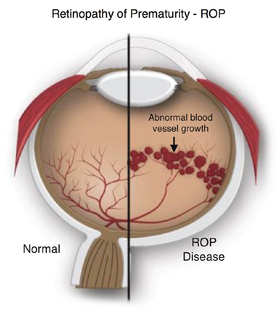
Prematurity can be challenging for children and families alike. The eyes are particularly susceptible to problems immediately after birth and for the rest of your child’s life. Retinopathy of Prematurity (ROP) is a potentially blinding eye disease that affects premature infants who have abnormal blood vessel development in the retina, the light-sensitive area of the eye where images focus.
Causes of Retinopathy of Prematurity
Blood vessels in the eye start developing in utero and if a child is born before that process is complete, there will be a part of the retina that does not have blood flowing to it. Blood vessels swell and overgrow in the light-sensitive layer of nerves in the retina at the back of the eye. When the condition advances, the abnormal retinal vessels extend into the jellylike substance (vitreous) that fills the center of the eye. Bleeding from these vessels may scar the retina and stress its attachment to the back of the eye, which can cause partial or complete retinal detachment and potential blindness.
Symptoms of Retinopathy of Prematurity
Visual symptoms of retinopathy of prematurity may include:
- Abnormal movement of the eyes
- Strabismus, or crossed eyes
- Myopia, or nearsightedness
- Pupils that are white
Who is at Risk for Retinopathy of Prematurity
ROP mostly strikes preemies who weigh 2¾ pounds (1250 grams) or less, those who are born before 31 weeks of gestation, or about 7 to 11 weeks before the normal gestation period. The disorder usually develops in both eyes and is among the most common causes of childhood vision loss. Although most cases of ROP resolve on their own, about 1 in 10 are severe enough to need medical intervention to prevent blindness.
In addition to a premature birth, other risk factors for developing retinopathy of prematurity may include the following:
- Anemia
- Medication
- Oxygen levels
- Blood transfusion
- Difficulty breathing
- Respiratory distress
- Health
- Low birth weight
Once your child has cleared the 50-week postmenstrual date, their risk for retinopathy of prematurity is quite low. However, your child may still be at risk for other eye problems as they get older including high myopia, or nearsightedness, as well as retinal tears or detachments in later life.
Eye Screenings for Preterm Babies
During pregnancy, blood vessels grow from the center of a developing baby’s retina 16 weeks into the mother’s pregnancy. They then branch outward and reach the edges of the retina between 8 months into the pregnancy and full term. In babies born early, normal retinal vessel growth may be disrupted, and abnormal vessels can develop which can cause leaking and bleeding into the eye. ROP has no signs or symptoms when it first develops in a newborn and the only way to detect it is through an eye exam by an eye doctor.
Shortly following birth doctors screen preterm babies for premature related illnesses, including ROP. Examination by the pediatric ophthalmologist may take place in a hospital’s nursery or neonatal intensive care.
What to Expect During An Examination
During the examination, the doctor will dilate your infant’s pupils with special drops. ROP may present at birth, or several weeks later. As such, premature infants at risk for ROP should have eyes examinations by an eye specialist a few weeks after being born. Infants born at less than 3.3 pounds (1500 grams) and before 31 weeks should routinely receive eye exams to check for the onset of ROP.
Vigilant monitoring is the key to ensuring the optimal outcome for your child and if early signs of retinopathy of prematurity present, laser treatment can slow the process in most patients. Surgery can also help in severe cases. These have both helped to decrease your child’s chances of severe vision loss in later life. Our eye care specialists will discuss how often to follow your child which ensures that they detect any early changes of retinopathy of prematurity.
For low-risk infants, it’s recommended to follow up every 2 weeks until 50 weeks of postmenstrual age, or 10 weeks after the original due date. Higher risk infants would need more frequent follow-up, either weekly or twice weekly.
Besides posing the alarming risk of possible blindness, ROP relates to vision irregularities later in life. An infant with ROP is more likely to develop certain eye problems as they grow, such as:
- Nearsightedness
- Lazy eye (amblyopia)
- Crossed or misaligned eyes (strabismus)
- Glaucoma
- Retinal detachment
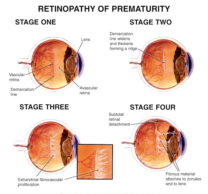
Stages of Retinopathy of Prematurity
The extent of abnormal blood vessel growth is categorized into five stages, based on the patient’s condition. Most of the infants diagnosed with ROP are diagnosed with mild cases, which includes some abnormal vessel growth but does not tend to cause permanent damage. More severe cases that involve significant growth of abnormal blood vessels may lead to retinal detachment and eventually cause vision loss or even blindness.
The stages of retinopathy of prematurity are classified as follows:
- Stage I: Mild, abnormal blood vessel growth. The disease usually resolves on its own without further treatment.
- Stage II: Moderate, abnormal blood vessel growth. The disease usually resolves on its own without further treatment.
- Stage III: Severely, abnormal blood vessel growth. The disease can resolve on its own but may require treatment depending on the degree of severity of the disease.
- Stage IV: Severely abnormal blood vessel growth and a retina that is partially detached that requires treatment.
- Stage V: The retina is completely detached and requires treatment to avoid total loss of vision.
Complications of Retinopathy of Prematurity
Retinopathy of prematurity can lead to a higher risk of developing the following conditions later on in life:
- Retinal detachment
- Myopia, or nearsightedness
- Strabismus, or crossed eyes
- Amblyopia, or lazy eye
- Glaucoma
- Blindness
Treatment of Retinopathy of Prematurity
In its early stages, many cases of retinopathy of prematurity do not require treatment. If the condition is advanced, laser therapy and cryotherapy are most often effective in destroying the peripheral areas of the retina to slow or reverse the growth of abnormal blood vessels. Laser treatments are used to burn away the periphery of the retina, while cryotherapy uses extremely cold temperatures to freeze the periphery. The side effects of either procedure includes a loss of peripheral vision, or side vision.
Additional treatment options for the advanced stages of ROP may include:
- Scleral buckle procedure
- Vitrectomy
Both of these surgical procedures may involve long-term side effects. Regular monitoring will also be performed to reduce the risk of vision loss and ensure that the patient’s eyes remain healthy and develop normally.
Contact SightMD today to schedule an appointment with one of our doctors to discuss your child’s vision health at one of our convenient locations!

Back-to-School Vision Checks: How Early Screening Sets Kids Up for Learning Success
The Hidden Key to School Readiness As August winds down, parents rush to stock up on backpacks, pencils, and…

Protecting Your Child’s Eyes This School Year
Why Eye Safety Matters During Back-to-School Season As the back-to-school season approaches, parents often prioritize pencils, backpacks, and lunchboxes….

August Is Children’s Eye Health & Safety Month: Spotting, Preventing & Treating Vision Issues
August marks Children’s Eye Health & Safety Month, a national observance dedicated to protecting one of your child’s most…