Retinal Detachment
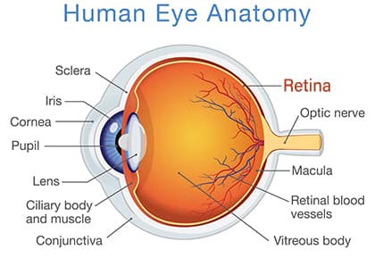
The retina is the light-sensitive tissue lining the back of the eye and plays a vital role in your ability to perceive images. There are several conditions that can permanently impair vision including retinal tears and retinal detachments. Early detection through regular eye examinations is a successful key in treating retinal tears and detachments helping your physician determine the appropriate treatment.
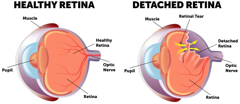
What’s a Retinal Tear or Detachment?
After the delicate tissue of the retina becomes torn, fluid can leak under the retina and lift it up eventually causing a total detachment. The longer this layer of tissue is separated from the blood vessels that supply it with oxygen and nutrients, the greater your risk of suffering permanent vision loss. Retinal detachment should be treated as a medical emergency as it often requires surgery to correct.
When to See an Eye Doctor
If not treated immediately, a retinal detachment can cause permanent vision loss. Anyone experiencing these symptoms of a retinal detachment should call the office immediately:
- The appearance of a curtain over the field of vision.
- Seeing light flashes
- Wavy or watery vision
- A sudden decrease in vision
- A sudden increase in the number of floaters in the field of vision
Who is Most at Risk for Retinal Detachment?
- Those who are very nearsighted
- The elderly
- People with a family history of retinal detachment
- Those who have had cataract surgery
- Patients with diabetes or other eye disorders
How is Retinal Detachment Diagnosed?
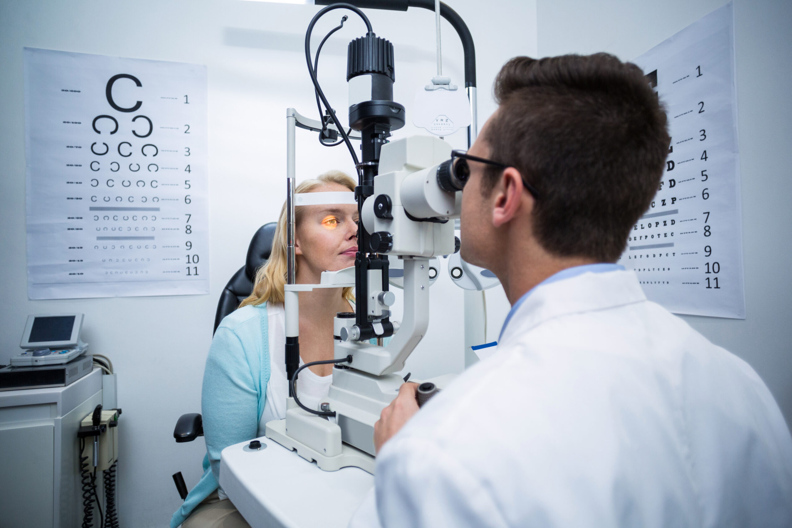
You need an eye exam to diagnose retinal detachment. Your eye care provider will use a dilated eye exam to check your retina. They’ll put eye drops in your eyes. The drops dilate, or widen, the pupil. After a few minutes, your provider can get a close look at the retina.
Your provider may recommend other tests after the dilated eye exam. These tests are noninvasive and won’t hurt. They help your provider see your retina clearly and in more detail:
- Optical coherence tomography (OCT): You get dilating eye drops for this imaging. Then you sit in front of the OCT machine. You rest your head on a support, so it stays still. The machine scans your eye but doesn’t touch it.
- Eye (ocular) ultrasound: You won’t need dilating drops for this scan, but your provider may use drops to numb your eyes so you won’t feel any discomfort. You sit in a chair and rest your head on a support, so it stays still. Your provider gently places an instrument against the front of your eye to scan it. Next, you sit with your eyes closed. Your provider puts gel on your eyelids. With your eyes closed, you move your eyeballs as your doctor scans them with the same instrument.
How is Retinal Detachment Treated?
Your eye care provider will discuss treatment options with you. You may need a combination of treatments for the best results. Treatments include:
Laser (Thermal) Therapy or Cryopexy (Freezing)
Sometimes, your provider will diagnose a retinal tear before the retina starts pulling away. Your provider uses a medical laser or a freezing tool to seal the tear. These devices create a scar that holds the retina in place.
Pneumatic Retinopexy
Your provider may recommend this approach if the detachment isn’t as extensive. After surgery, your provider will recommend that you keep your head still for a few days to promote healing. You also may be told not to lie on your back.
Scleral Buckle
During this procedure:
- Your provider surgically places a silicone band (buckle) around the eye.
- The band holds the retina in place and stays there permanently. You can’t see the band.
- The detached retina starts healing.
- Laser or cryopexy are used to seal the tear.
Vitrectomy
During a vitrectomy, your provider:
- Surgically removes the vitreous.
- Places a bubble of air, gas or oil in the eye to push the retina back in place
If your provider uses an oil bubble, you’ll have it removed a few months later. Gas and air bubbles get reabsorbed. If you have a gas bubble, you may have to avoid activities at certain altitudes. The altitude change can increase the size of the gas bubble and the pressure in your eye. You’ll have to avoid flying and traveling to high altitudes. Your provider will tell you when you can start these activities again.
Can I Prevent Retinal Detachment?
You can’t prevent retinal detachment, but you can take steps to lower your risk:
- Get regular eye care: Eye exams protect your eye health. If you have nearsightedness, eye exams are especially important. Myopia makes you more prone to retinal detachment. Your eye care provider should include dilated exams to find small retinal tears.
- Stay safe: Use safety goggles or other protection for your eyes when playing sports or doing other risky activities.
- Get prompt treatment: If you notice detached retina symptoms, see your eye care provider right away or go to the emergency room.
Contact SightMD today to schedule an appointment with one of our doctors to discuss your vision health at one of our convenient locations!

Flashes and Floaters: When Are They an Eye Emergency?
Changes in vision, such as flashes of light or floaters, can be unsettling. While these symptoms are often benign,…
Understanding Laser Surgery for Retinal Tear
What to Expect During Recovery after Laser Surgery for Retinal Tear Laser surgery for a retinal tear is a…

Understanding what Age-Related Macular Degeneration is
Age-related macular degeneration (AMD) is the most common cause of vision loss in individuals over the age of 50….